


Sign up for CNN’s Wonder Theory science newsletter. Explore the universe with news about fascinating discoveries, scientific breakthroughs and more.
An extinct ribbon-shaped sea creature the size of a human thumb was one of the first animals to develop a precursor to a backbone. Scientists recently identified the animal’s nerve cord using an upside-down twist. They turned their fossils upside down.
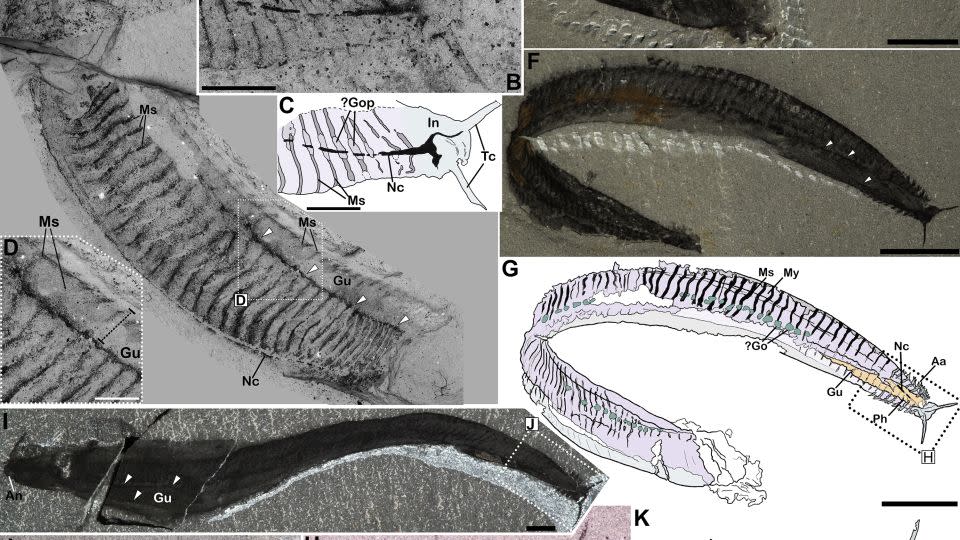
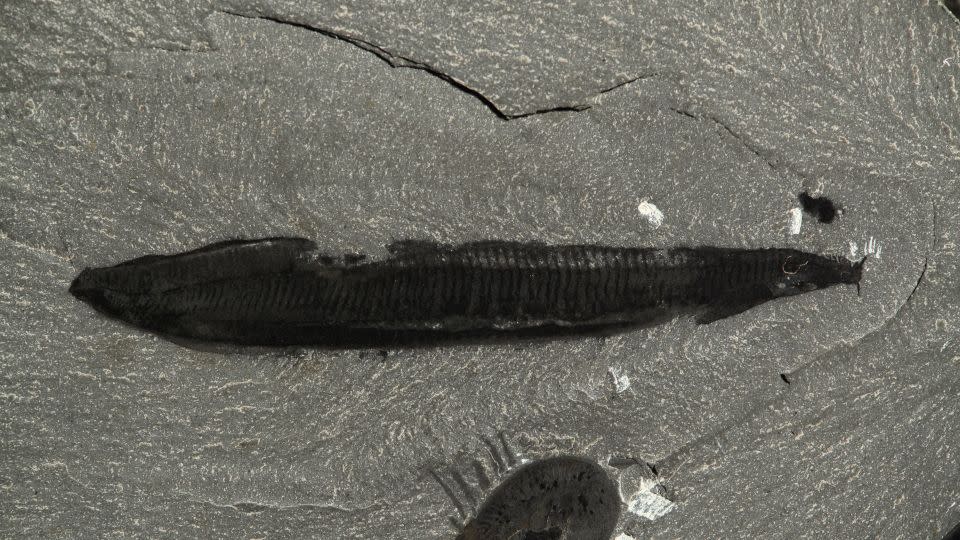
Paleontologist Charles Doolittle Wolcott first found Pikaia fossils in the Burgess Shale deposits of British Columbia, dated to 508 million years ago, and described them in a 1911 treatise. The animal was about 4 centimeters long on average and had a flattened body and sinuous and a tiny head, with two tentacles at the tip and fringed with external gills. These were originally thought to be rudimentary legs, so the animal was positioned with these structures facing downwards.
In 2012, after decades of studying Pikaia fossils, researchers described its internal structures fossilized in great detail. They identified a long thread near the belly as a blood vessel and named a 3D sausage-shaped structure running down the animal’s back as a dorsal organ, possibly used for internal support, although such an organ would be anatomically different from anything seen in fossils. or in living beings. animals.
However, a recent analysis of the Pikaia fossils by another team of scientists, published June 11 in the journal Current Biologyoverturned this view and all other previous studies on Pikaia.
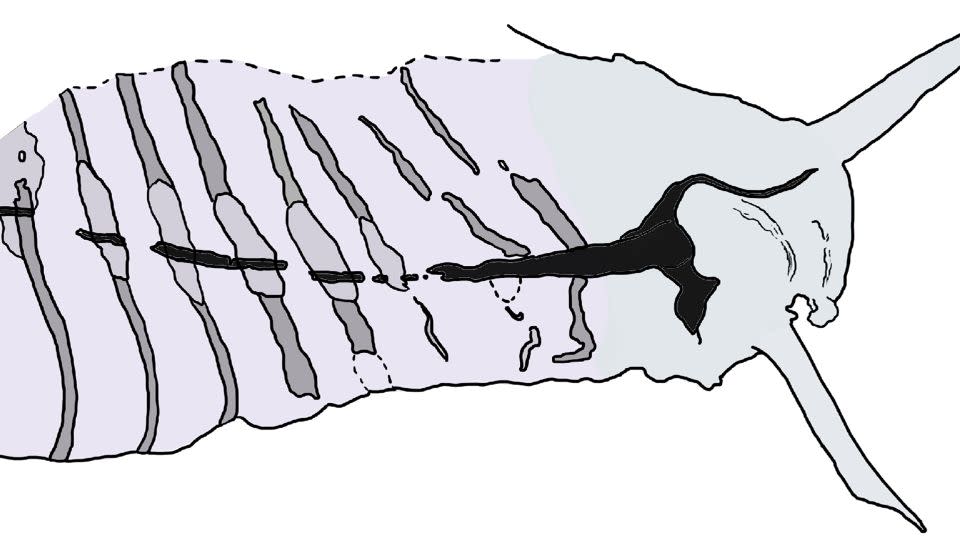
According to the researchers, previous anatomical interpretations positioned the animal wrong side up. The so-called dorsal organ was actually located in the belly and was Pikaia’s intestine. The supposed blood vessel was a dorsal nerve cord, a feature associated with the group of animals known as chordates, in the phylum Chordata.
All chordates, such as vertebrates, eel-like lancelets, and tunicates or sea squirts, at some point in their lives have a flexible, rod-shaped nerve structure called notochord on your back. A dorsal tubular nerve cord is also a characteristic of chordates.
Pikaia was initially thought to be a worm, but was later upgraded to an early type of chordate, based on features such as the shape of certain muscles and the position of its anus. But experts weren’t sure where exactly Pikaia belonged in the chordate family tree.
With the description of a dorsal nerve cord, Pikaia can now be considered part of the fundamental lineage of all chordates, although it has no direct descendants who are alive today, the study authors reported.
Flipping Pikaia “clarifies things a lot”, says evolutionary biologist Dr., clinical professor at the University of Idaho. Mallatt, who was not involved in the new research, published a paper on Pikaia in 2013working from the established body position (and upside down).
In retrospect, the truth was “hiding in plain sight,” and the reversal in orientation resolves questions about why Pikaia’s putative blood vessel and dorsal structure conflicted with established anatomical features in other chordates, Mallatt said.
“Pikaia suddenly became a lot less weird,” he said.
New guidance
The reassessment of the path followed by Pikaia came years ago with a co-author of the new study, Dr.professor of macroevolution at the University of Bristol in the United Kingdom, said the study’s lead author Giovanni Mussiniresearcher and doctoral candidate in the department of Earth sciences at the University of Cambridge, in the United Kingdom.
There were a number of reasons to revisit previous interpretations of the fossils, Mussini told CNN. On the one hand, there was the enigma of what scientists believed to be the position of the dorsal organ. Its location – near what was supposed to be Pikaia’s back – apparently ruled out the possibility that the organ could be an intestine.
However, when Pikaia was turned upside down, the organ’s location and characteristics made more sense anatomically. It enlarged and extended into the animal’s pharynx, the region of the throat where the intestine normally connects to the mouth. Its 3D status could be explained by the presence of chemically reactive tissues – characteristic of a gut. In other Burgess Shale fossils, abundant ions and reactive compounds that are normally found in intestinal tissue cause the digestive structures to mineralize more quickly than the rest of the body and thus retain more of their original forms. The structures inside Pikaia’s organ were possibly remnants of swallowed food, according to the study.
In an inverted Pikaia, the external gills that previously pointed downwards were now angled upwards, as are the external gills in modern mudskippers and axolotls.
Flipping Pikaia also changed the orientation of the muscle groups that group together in a wave formation. These muscles, called myomeres, are a fundamental feature of vertebrates. In Pikaia’s new position, the strongest point of flexion of these muscles is along the back, which also applies to the arrangement of the myomeres in other animals with spinal columns.
“This makes Pikaia’s movement consistent with what we see in modern chordates,” Mussini said.
Finding the courage
Pikaia’s putative blood vessel was also anatomically intriguing, as it lacked the branches normally found in vertebrate blood vessels.
“It’s a single line that runs through most of the body to the head, where it forks into two threads that form the tentacles,” Mussini said.
An important part of recognizing the structure as a nerve cord was the fossilized nervous systems in other Cambrian Period animals (541 million to 485.4 million years ago) that were discovered in the last decade, Mussini added.
“We have a better understanding of how nerve cords and other tissues fossilize because we have been lucky enough to find some Cambrian nervous systems preserved in other deposits,” he said, “mostly from Chinese fossils that have come to light in the last few years.”
Many of these fossils were arthropods – invertebrates with exoskeletons – with living relatives such as insects, arachnids and crustaceans; Comparing the fossils to modern arthropods helped paleontologists identify preserved internal tissues. One example is a fossil specimen of the Cambrian arthropod Mollisonia, which showed brain organization comparable to that of living spiders, scorpions and horseshoe crabs, Mussini said.
Although there are no living analogues for Pikaia, data from arthropod fossils have given scientists a more detailed frame of reference for Pikaia’s nerve cord. Like other fossilized nerve tissue, Pikaia’s nerve cord was dark, rich in carbon, and relatively fragile compared to other fossilized tissue.
This dorsal nerve cord solidifies Pikaia’s status as a chordate, placing it “pretty much at the base of what we would consider traditional chordates,” Mallatt said.
Much about Pikaia’s anatomy remains a mystery, but looking at it from a new angle could offer new insights into its intriguing range of features, Mussini said.
“Many of these details have only come to light in the last 10 or 12 years,” Mussini added. “The authors of the 2012 paper can certainly be forgiven for not bringing these details into the conversation, because it is a work in progress.”
Correction: A previous version of this story gave an incorrect average length of Pikaia.
Clarification: This story has been updated to reflect that the dorsal nerve cord is a feature associated with chordates and the notochord is a feature of chordates that is distinct from the dorsal tubular nerve cord.
Mindy Weisberger is a science writer and media producer whose work has appeared in Live Science, Scientific American, and How It Works.
For more news and newsletters from CNN, create an account at CNN.com


/cdn.vox-cdn.com/uploads/chorus_asset/file/23986638/acastro_STK092_02.jpg?w=150&resize=150,150&ssl=1 "YouTube is canceling Premium subscriptions with fake locations")